Research
Current Projects
Modeling of Cortical Color Responses
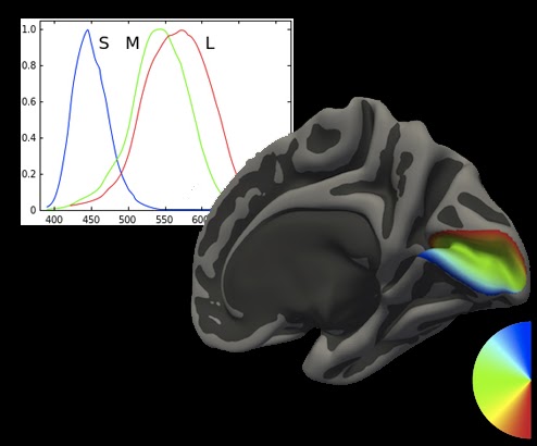
Examine the representation of color stimuli in early visual cortex through the use of functional magnetic resonance imaging (fMRI). The goal of this project is to present subjects with targeted light spectra that selectively stimulates the photoreceptors in order to model the cortical sensitivity to various color directions. We developed a model of the BOLD fMRI response to these chromatic stimuli based on elliptical isoresponse contours. We call this model the quadratic color model (QCM). Using the QCM parameterized fits to the fMRI time course, we examine how these parameters vary based on factors such as eccentricity and visual area. For more information, see our paper in eLife.
Examine the representation of color stimuli in early visual cortex through the use of functional magnetic resonance imaging (fMRI). The goal of this project is to present subjects with targeted light spectra that selectively stimulates the photoreceptors in order to model the cortical sensitivity to various color directions. We developed a model of the BOLD fMRI response to these chromatic stimuli based on elliptical isoresponse contours. We call this model the quadratic color model (QCM). Using the QCM parameterized fits to the fMRI time course, we examine how these parameters vary based on factors such as eccentricity and visual area. For more information, see our paper in eLife.
Temporal Dynamics of Color Tracking
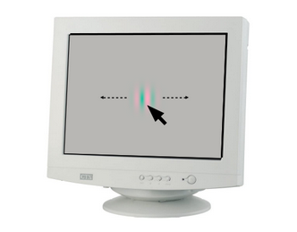
Examining the timescale of color processing. This is done by using a novel psychophysics experiment in which participants attempts to track the position of a patch made up of various colors that jump around on a computer monitor. From this tracking data, temporal response functions are estimated for each color that was shown to a participant (color specifically refers to the ratio of L- and S- cone activity). These response functions allow us to determine properties such as the temporal delay which are needed to understand how different colors are processed at different timescales by the visual system.
Examining the timescale of color processing. This is done by using a novel psychophysics experiment in which participants attempts to track the position of a patch made up of various colors that jump around on a computer monitor. From this tracking data, temporal response functions are estimated for each color that was shown to a participant (color specifically refers to the ratio of L- and S- cone activity). These response functions allow us to determine properties such as the temporal delay which are needed to understand how different colors are processed at different timescales by the visual system.
Past Projects
Predictive Atlas of High Level Visual Areas
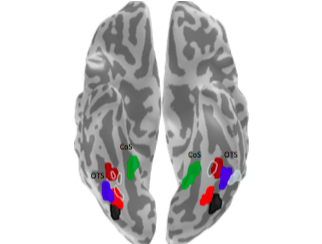
Category selective functional regions of interest (fROI) were used to create maximum probability maps (MPM) of ventral temporal cortex. Using cortex based alignment, I registered these MPM fROIs to an average cortical surface and used a leave one out analysis that allowed for the quantification of the spatial predictability of these fROIs.
Category selective functional regions of interest (fROI) were used to create maximum probability maps (MPM) of ventral temporal cortex. Using cortex based alignment, I registered these MPM fROIs to an average cortical surface and used a leave one out analysis that allowed for the quantification of the spatial predictability of these fROIs.
Function and Cytoarchitecture
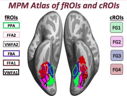
Created cortical mesh reconstructions of post-mortem human brains in FreeSurfer based on hand segmentations. The surfaces were used to create maximum probability maps (MPM) of cytoarchitechtonic regions of interest (cROI) in the fusiform gyrus. I created a pipeline using the cortex based alignment tools of FreeSurfer to register these MPM cROIs to individual living subjects. For the first time, this allowed the quantification of proportion overlap of category selective regions of VTC and its cellular composition.
Created cortical mesh reconstructions of post-mortem human brains in FreeSurfer based on hand segmentations. The surfaces were used to create maximum probability maps (MPM) of cytoarchitechtonic regions of interest (cROI) in the fusiform gyrus. I created a pipeline using the cortex based alignment tools of FreeSurfer to register these MPM cROIs to individual living subjects. For the first time, this allowed the quantification of proportion overlap of category selective regions of VTC and its cellular composition.

Development of Perception
A longitudinal research study examining human high-level visual cortex in children (5-12 years old) and adults. From this study, we found growth of tissue within portions of ventral temporal cortex, challenging the idea that cortex is either quiescent or pruned after early childhood. This tissue growth within face-selective cortex, is correlated with increases in the functional selectivity of face-selective regions and linked to improvements in recognition memory ability well into adulthood. To do this study, we combined quantitative magnetic resonance imaging techniques with functional MRI and behavioral measurements in single-subject analyses. We have also linked these measures with ex vivo quantifications of cytoarchitecture.
A longitudinal research study examining human high-level visual cortex in children (5-12 years old) and adults. From this study, we found growth of tissue within portions of ventral temporal cortex, challenging the idea that cortex is either quiescent or pruned after early childhood. This tissue growth within face-selective cortex, is correlated with increases in the functional selectivity of face-selective regions and linked to improvements in recognition memory ability well into adulthood. To do this study, we combined quantitative magnetic resonance imaging techniques with functional MRI and behavioral measurements in single-subject analyses. We have also linked these measures with ex vivo quantifications of cytoarchitecture.
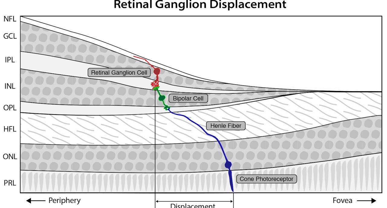
Modeling of Retinal Ganglion Cell Displacement
Model the lateral displacement between cones and retinal ganglion cells in the human retina. Correcting for this spatial displacement is needed to relate measurements of ganglion cells to measurements of cones, perception, and cortex. We have developed a spatial model that solves for RGC displacement at any arbitrary retinal position. The goal of this model is to use individual subject data to link measurements of cone and RGC density to visual function and cortical organization.
Model the lateral displacement between cones and retinal ganglion cells in the human retina. Correcting for this spatial displacement is needed to relate measurements of ganglion cells to measurements of cones, perception, and cortex. We have developed a spatial model that solves for RGC displacement at any arbitrary retinal position. The goal of this model is to use individual subject data to link measurements of cone and RGC density to visual function and cortical organization.